Low back pain: causes and diagnosis
4 min read
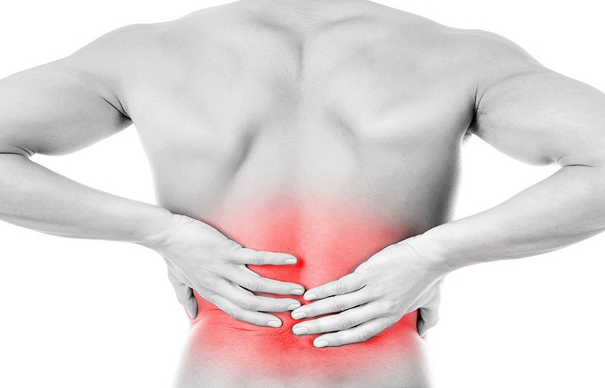
The pain lumbar or lower back pain is the pain located between the subcostal region and the gluteal fold, irradiated or referred many times to the sacroiliac region or thighs. It is accompanied by tension, spasm, or muscle stiffness, sometimes radiating down the leg.
Low back pain is a highly prevalent problem in the population (80% of people have had low back pain at some point in their lives) due to its economic and social repercussions. It is one of the first causes of sick leave due to incapacity for work in developed countries.
It is essential to differentiate between acute or chronic low back pain, trying through the medical history to reach the cause of this pain. 85% of the time, the pain is nonspecific, without the presence of a defined pathological lesion. It is diagnosed as “low back pain, lumbago, impingement ” a term that means: low back pain, and, to physiotherapists, it does not contribute anything, since the fact that the patient comes with that low back pain already gives the same information.
In contrast, specific low back pain is found after a defined injury such as an infection, osteoporosis, or fracture.
Low back pain diagnosis
As in any medical history, before performing a physical examination, the Doctor must carry out an anamnesis that leads to the most accurate diagnosis possible to treat the patient.
- The appearance of pain, acute or chronic? How did it appear? It is essential to know if the patient has suffered a blow or fall, if it has been as a result of physical activity (it gives rise to a possible fracture, contusion, or post-traumatic muscle spasm) or if it has appeared suddenly but without suffering any blow ( possible infection, osteoporosis, herniated disc).
- On the contrary, if the pain seems gradual and continues over time, with no apparent associated cause, Then it must be investigated further into the type of pain, location, and intensity.
- Location: Is it in a localized spot? Does it radiate, or does it refer to other areas of the body? Is it located in a bar in the lumbar region? This can give the information about pain with nerve radiation (for example, sciatica), presence of myofascial trigger points (possible points in the lumbar area that give pain referred to the gluteal region or vice versa, that the origin of the pain is the gluteal or abdominal area but refer pain to the lower back), visceral pain radiating to the lower back (for example pancreatic or pelvic)
- Intensity and type of pain: Assess with intensity scales, for example, VAS (visual analog scale) or Scale from 0 to 10, and their variations. What factors increase pain? (postures, activity, time of day) What factors decrease it? (medication, analgesic postures, stretching, physiotherapy).
- Mechanical pain is related to effort, movement or posture, calm with rest, and analgesic postures. The inflammatory pain is gradual, progressive, continuous, nocturnal, unrelated to action or posture: neoplasms, infections. The pain of spinal canal stenosis produces claudication when walking with sensory and motor alterations, weakness.
It is essential to differentiate the type of pain since visceral or infectious pain, in general, does not stop or change with relaxed postures or with rest, so after anamnesis and exploration, and if there is evidence of any systemic disease refer to a doctor, fascial pain especially during the night awake since the system is “uncomfortable” maintaining the same posture for a long time, pain of muscle origin decreases with rest, etc.
The Doctor must take into account all these elements and then proceed with the physical examination:
- Observation: In a dynamic statement, A doctor will see the patient’s movements, if they are free, if he is afraid, if he presents any antalgic posture or if he has a feeling of discomfort when moving.
- Palpation: pain on palpation in the area referred to by the patient? Discomfort in other points that refer pain to the lumbar region? (presence of PGM) increased muscle tone? Restricted mobility of skin and subcutaneous cellular tissue? Pain in spinous processes? Anteriorization or posteriorization of lumbar transverse, iliac?)
- Mobilization, muscle strength and specific tests (muscle strength tests of abdominal, lumbar, multifid, gluteal, hamstring, iliac psoas), extensibility tests, and specific tests (Lasègue test, Schober test, Gillet.)
- Complementary tests: The presence of diagnostic tests is interesting if a doctor suspect infections, herniated disc, spondylolisthesis, etc., to guide our treatment or refer to another professional. Although the doctor does not find a specific injury in most cases, so the patient is treated with anti-inflammatories and sent home, then this is where our assessment and treatment arrives.


